


General Health
Can You See a Private GP While Registered with the NHS?
We get asked all of the time, “Is it OK that I have an NHS GP?” It absolutely is! It’s


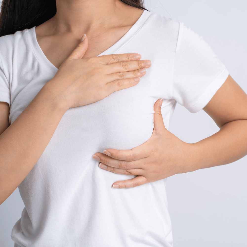
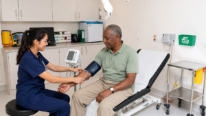
Preparing for a mammogram involves wearing a two-piece outfit and avoiding deodorants or lotions on your chest. Bring past mammogram records and be ready to discuss any breast symptoms. The procedure may be briefly uncomfortable but is generally painless. Follow specific guidelines if an ultrasound is also scheduled.

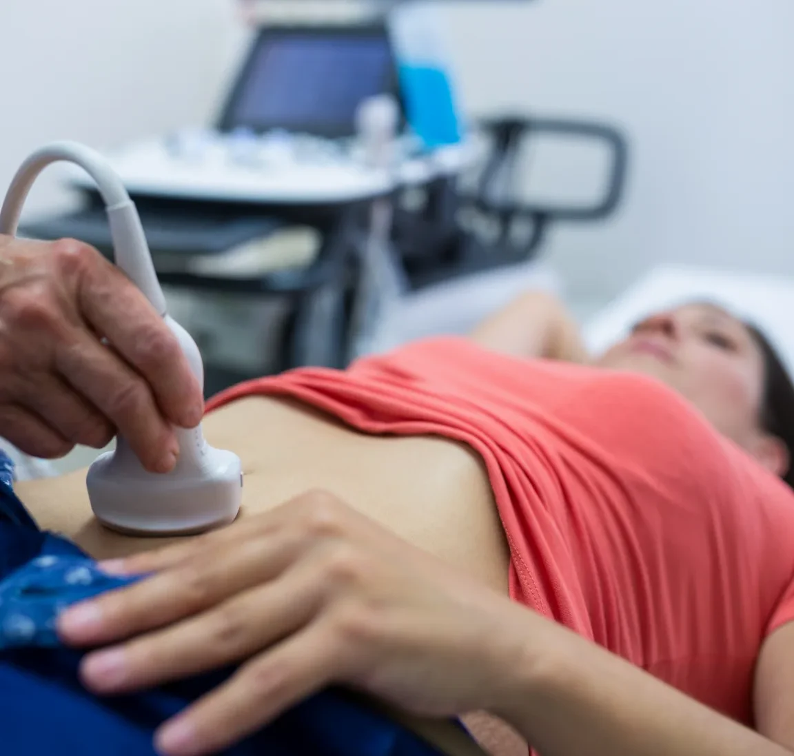
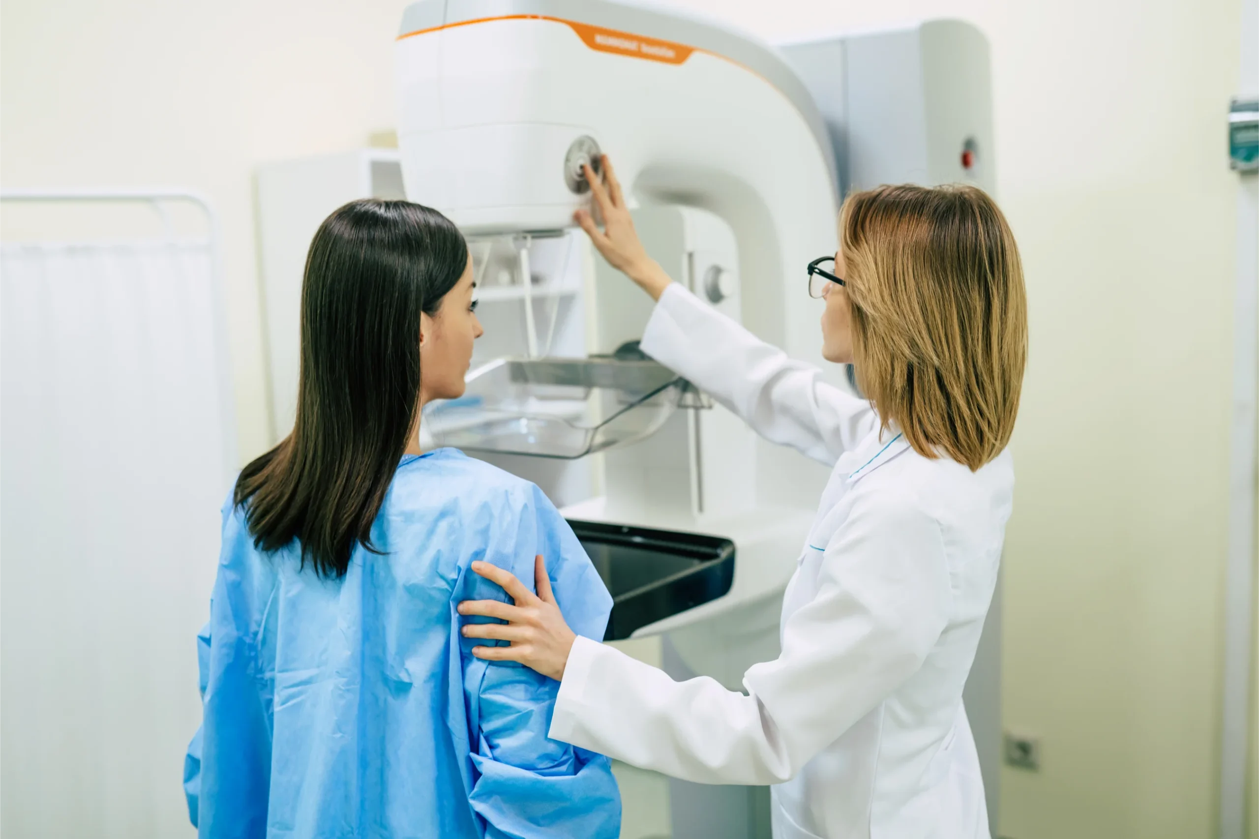
During a mammogram with X-Ray and ultrasound, you will wear a gown and have your breast gently compressed for X-Ray images, which may briefly cause discomfort but helps in imaging. If ultrasound is needed, gel is applied, and a transducer captures images with sound waves. The process takes about 30 minutes.

After a mammogram with X-Ray and ultrasound, you can resume normal activities immediately. Results are reviewed by your healthcare provider. Most mammograms show no signs of breast cancer, but further steps may be recommended if needed.
We get asked all of the time, “Is it OK that I have an NHS GP?” It absolutely is! It’s
Written by a medical professional: Alya Shakir. Medically reviewed by Dr Enam Abood. Many Londoners start the working week feeling
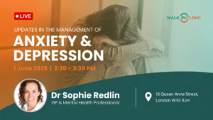
City Walk-in Clinic is pleased to announce an educational event that could be useful to the general public as well